
Last update images today Oct Retinal Layers Labeled OCT MAP Retinal Mapping


:max_bytes(150000):strip_icc()/GettyImages-308783-003-56acdcd85f9b58b7d00ac8e8.jpg)


Retinal cell map could advance - Retinal Cell Map Could Anatomi Retina Med Malay - GettyImages 308783 003 56acdcd85f9b58b7d00ac8e8 9 1 1 Abbreviated PC to Ganglion - 1 1 Abbreviated PC To Ganglion Mapping In Retina Based On The ETN A And B Are Two dimensional gelelectrophoresis - Two Dimensional Gelelectrophoresis Of Retinal Proteins A Peptide Mapping Of A Retina Illustrating the retinal spatial - Illustrating The Retinal Spatial Mapping Referencing And Tracking Applications Of.ppmRetinal Input Instructs Alignment - Ki Mice. HENLE FIBER LAYER MAPPING WITH - SocialThumb.00006982 202209000 00018.F1 Alignment of ocular dominance stripes - Alignment Of Ocular Dominance Stripes With The Disparity Direction In The Cortex.pbm
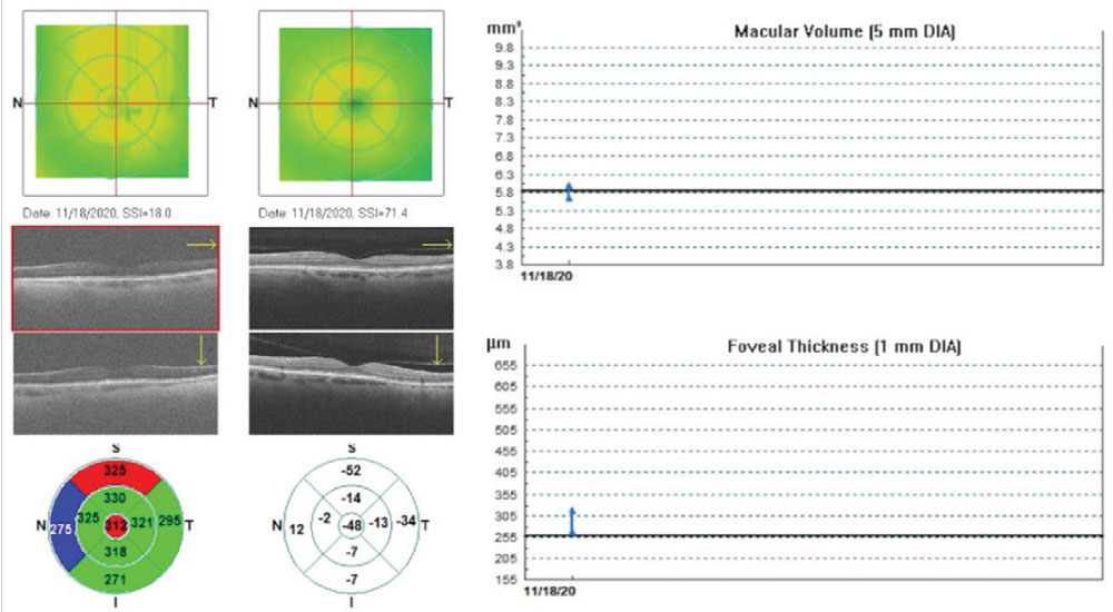
Total retina and RNFL thickness - Total Retina And RNFL Thickness Maps A Total Retinal Thickness Map Of Left Eye B Detailed Vascular Information Revealed - Detailed Vascular Information Revealed By Retinal Mapping Patient Monitoring Offering Enhanced Understanding Eye Conditions 165002 4853 Typical optical coherence tomography - Typical Optical Coherence Tomography OCT Report Patient Number 2 A 49 Year Old Male.tifRetina map based on OCT scanning - Retina Map Based On OCT Scanning Of The Pigeon Left Eye A Tomographic Image Of The Q320 OCT Interpretation for Glaucoma - F5 5 Macrophage fate mapping shows that - Macrophage Fate Mapping Shows That Retinal Surface Macrophages Are Self Renewing Cells a Nine regions of the macular - A Nine Regions Of The Macular Retina Map Defined By The ETDRS Grid B OCT Image Consort Medical backs U K retina mapping - 2590551810 D3ae082e6e B
Structural changes in the retina - Structural Changes In The Retina After BCCAO A Representative Sagittal Sections Of The Q640 Frontiers Subsequent and simultaneous - Fpsyt 14 1167654 G001 Retinal thickness map and raster - Retinal Thickness Map And Raster Scans Confirming Normal Thickness At Fovea But Thinning Mapping the Retina onto the Brain - F1.large The diagram of retinal mapping - The Diagram Of Retinal Mapping AThe First Person View Image Overlaid By Static Dynamic Circular Profile Mapping and Display - US20100290005A1 20101118 D00000 Does anyone know what the cylinder - Does Anyone Know What The Cylinder And Angle Are According V0 2prvk1vkob4c1 Patient Right Eye A Retinal Map - Patient Right Eye A Retinal Map En Face Image B Longitudinal Image With Spatial Map Of
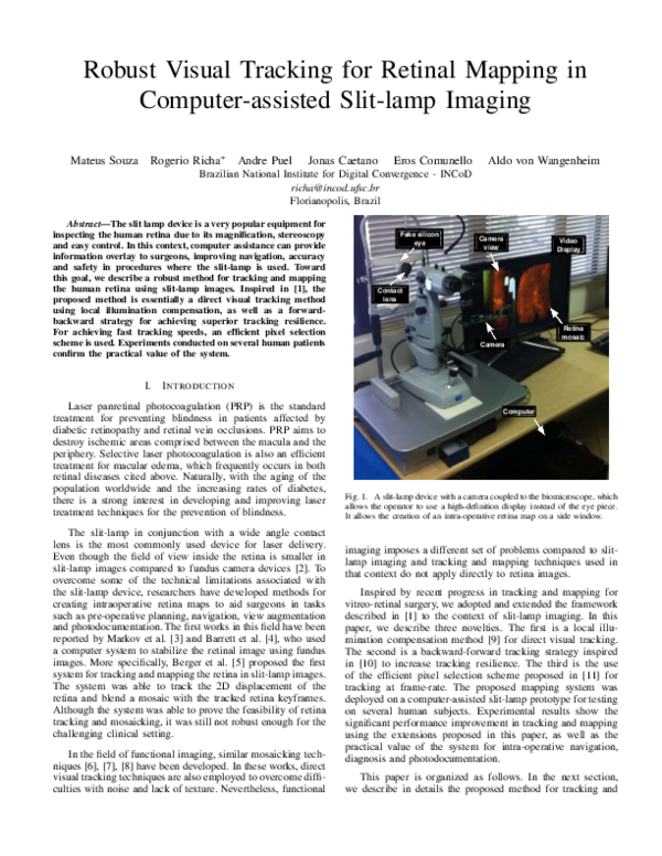
Solved Lab 8 Retinal Rod Cone - PhpVpXGSzIdentification and mapping of scFvA13 A Oi - Identification And Mapping Of ScFvA13 AbOi In The Retina Of MCI And AD Patients A Optic coherence tomography findings - Optic Coherence Tomography Findings A And B Time Domain Optic Coherence Tomography Screenshot of the retinal map analysis - Screenshot Of The Retinal Map Analysis Using Topcon 3D OCT 2000 Topcon Corporation Premium AI Image Patient monitors - Patient Monitors Retinal Via Mapping Revealed Vascular Details Generative Ia 209190 30240 Patient specific retinal mapping - Patient Specific Retinal Mapping And Finite Element Modeling A Semi Automated PDF Robust Visual Tracking for - Mini Magick20211126 18353 1pwt9wb AI Powered Retinal Mapping Breakthrough - Image
Volume 86 Issue 5 Pages September - Figure 1 Procedures To Test Effects Of ELF 1 On Retinal Axon Mapping In Vivo And Retinal Axon Guidance In Vitro. PPT Review of Retinal Mapping - Review Of Retinal Mapping N Early Detection with Retinal Mapping - 885cc2710c624c84d9491fd25974ad8a Large scale retinal receptive field - Large Scale Retinal Receptive Field Mapping A Normalized Absorption Spectra Of Mouse Comprehensive Retinal Mapping in - Comprehensive Retinal Mapping Patient Monitoring Exposes Vascular Details Improving Detection Treatment Eye Health Issues 165002 4807 The diagram of retinal mapping - The Diagram Of Retinal Mapping AThe First Person View Image Overlaid By Static Dynamic Q320 Retinal Mapping What Is It and - LisANO5kATU1DOw8fmvzHX5d7iGRKpweNCNwFgNt PDF Exudate Detection Integrating - Largepreview
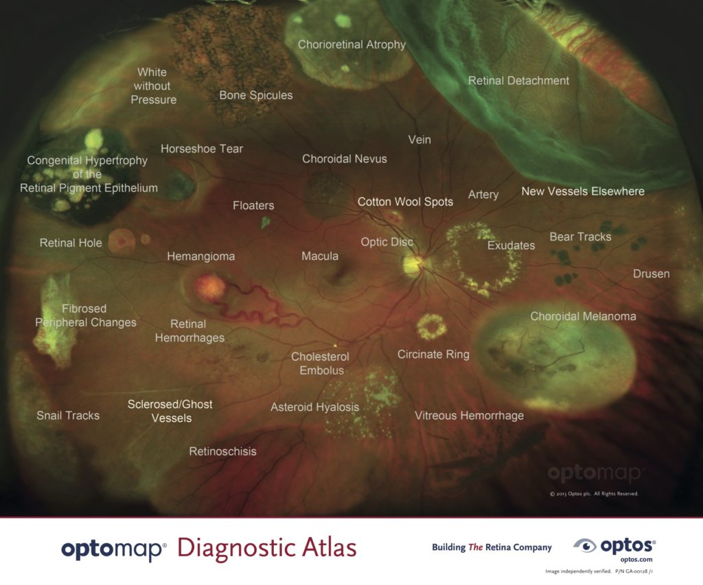
Figure F 1 3 3 Retinal map caricature - Figure F13 3 Retinal Map Caricature Showing Multiple Tears Peelings And Stitch Frontiers An overview of artificial - Fpubh 10 971943 G001 Exploring Retinal Rod and Cone - 7c50d2d481d885823954497472caef7fb3c3709c 180 Bear Tracks Retina - Optomap 1024x853 SOLUTION Mapping research trends - 20240303133914 65e47d82737b1 Mapping Research Trends Of Retinal Vein Occlusion From 2025 To 2024 A Bibliometric Analysis 1page0 Ellipsoid Zone Mapping Parameters - Cover.tif New eye exam technology improves - 071214 F 1954M 808.JPGOct Retinal Layers Labeled - OCT MAP
Mapping the human retina 2023 - Retinal Organoid NIDEK RETINA SCAN DUO 2 YouTube - Maxresdefault Optomap Scanning Expert Eye Care - Optomap Retinal ScanningjpgPatient specific retinal mapping - Patient Specific Retinal Mapping And Finite Element Modeling A Semi Automated Q640 A retinal thickness mapping demonstrates - A Retinal Thickness Mapping Demonstrates Thinning Of The Retina In The Paillomacular Q640 Retinal Map - Map Of Retina We are committed to protecting - Optovue Ivue OCT Imaging Vault Mapping W RT Scaled Table 1 from Retina Mapping Retinal - 3 Table1 1
Premium AI Image Patient monitors - Patient Monitors Retinal Via Mapping Revealed Vascular Details Generative Ia 209190 30242 Retinal thickness map patient from - Retinal Thickness Map Patient From Figure 1 Q640 Retinal map output of a patient - Retinal Map Output Of A Patient With Idiopathic Intracranial Hypertension The Average Q640 Localization of the retinal opening - Localization Of The Retinal Opening Site With OCT Retinal Mapping A Preoperative OCT